a meta-analysis of neuroimaging evidence of visual mental imagery
What do we know about the neural basis of visual mental imagery from past fMRI studies?
Abstract
The dominant neural model of visual mental imagery (VMI) stipulates that memories from the medial temporal lobe acquire sensory features in early visual areas. However, neurological patients with damage restricted to the occipital cortex typically show perfectly vivid VMI, while more anterior damages extending into the temporal lobe, especially in the left hemisphere, often cause VMI impairments. Here we present two major results reconciling neuroimaging findings in neurotypical subjects with the performance of brain-damaged patients: (1) A large-scale meta-analysis of 46 fMRI studies, of which 27 investigated specifically visual mental imagery, revealed that VMI engages fronto-parietal networks and a well-delimited region in the left fusiform gyrus. (2) A Bayesian analysis showed no evidence for imagery-related activity in early visual cortices. We propose a revised neural model of VMI that draws inspiration from recent cytoarchitectonic and lesion studies, whereby fronto-parietal networks initiate, modulate, and maintain activity in a core temporal network centered on the fusiform imagery node, a high-level visual region in the left fusiform gyrus.
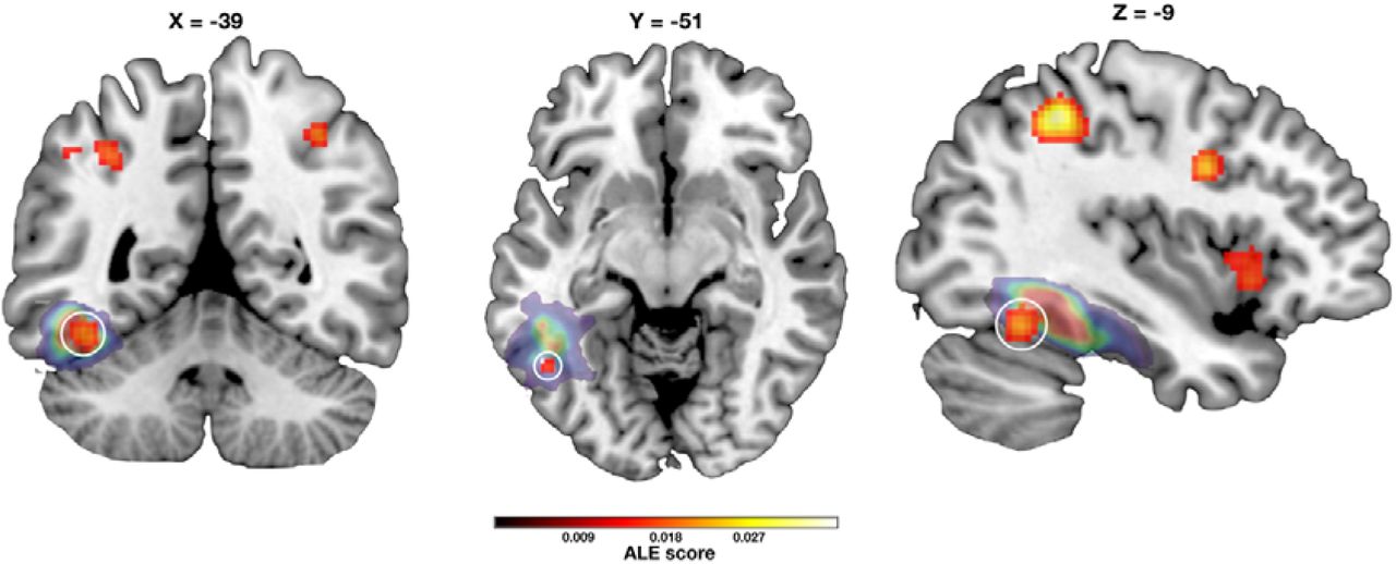
(MNI coordinates: x=-42, y=-54, z=-18)
Find our paper here.
Enjoy Reading This Article?
Here are some more articles you might like to read next: